
What set O'Shaughnessy apart was
the difficulty of the problems he sought to solve,
the vigor with which they were pursued,
and the unique solutions he proposed.
ORWELL DOC O'SHAUGHNESSY HEART OMEN
Up to then, all the focus regarding revascularization of tissue
had been concentrated on vessel surgery
(unblocking, bypassing, or modulating through the autonomic nervous system).
Cardiac ischemia, however, remained a problem.
O'Shaughnessy spawned the idea
of bringing in tissue with excellent vascularization abilities
and devised the route to get it to the ischemic heart.
He chose omentum.
To Orwell Today,
Dear Ms. Jura,
Attached find a copy of a little paper that appeared in the esteemed Annals of Thoracic Surgery on February the 1st, 2015.


Should you find it useful, you are more than welcome to refer to it in your Blog.
Kind regards,
Jan J van Wingerden, FCS
Greetings Jan van Wingerden,
Congratulations on the publication of your fascinating paper -- and thanks so much for sending a copy to share with ORWELL TODAY readers. I recall our previous discussion during research mode. See ORWELL DOC O'SHAUGHNESSY GIANT IN FIELD
Below, to enhance understanding -- especially for those not familiar with medical terms -- I've transcribed and highlighted excerpts, inserted images and added a glossary -- to keep the story flowing like a river running through it.
All the best,
Jackie Jura
OUR SURGICAL HERITAGE
Sternotomy and Intrathoracic Omentum: Two Procedures, Two Innovators,
and the River That Runs Through It -- A Brief History
by Jan J van Wingerden, MBChB (UP), FCS (SA)
Academic Medical Center, University of Amsterdam, Amsterdam, The Netherlands
This is a small tribute to the monumental contributions of the early pioneers who introduced 2 invaluable thoracic procedures; an exposure through which the mediastinum and the heart could be safely approached, the median sternotomy, and an approach and method to revascularize an intrathoracic organ by transferring the omentum into the chest. The origin, rediscovery, and application of these procedures are strangely linked. With the renewed interest in variations of the median sternotomy and the effectiveness of using the intrathoracic omentum, the legacy of the original innovators lives on.
There is some learned debate about whether the dawn of cardiac surgery was heralded by the deliberate opening of the pericardium to release the entrapped heart from the dark, red fluid holding it captive, or whether the Dark Ages preceding cardiac surgery finally ended with the successful closure of the ruptured pericardium or myocardium, all the result of a penetrating injury of the chest. Be that as it may, these were courageous attempts and interestingly essayed. The new dawn certainly rose with the news of the long-term survival of a patient after successful cardiac repair.

In 1895, H.C. Dalton of St Louis, Missouri, reported on the survival of his patient whose cardiac stab wound he had explored 4 years earlier. Only the pericardial sac was found punctured. It was repaired with a continuous catgut suture.... The reluctance of all surgeons of the time to suture the myocardium has to be seen in context. It was based on the, albeit unsubstantiated, then current belief that the slightest injury to the myocardium, even that of a suturing needle, would result in immediate cardiac arrest. Thus the ethical dilemma was this: would it be justifiable to risk certain death due to cardiac arrest with the attempt at suturing the myocardium in a patient who was, up to the point of suturing, still alive. The dilemma was resolved [1897] when Ludwig Rehn of Frankfurt am Main, Germany, presented the first surviving case of myocardial suturing after a stab wound the same year (1897).... He expounded on and justified his act to his colleagues...stating that the risk of dying after stab to the heart is more probably due to uncontrolled hemorrhage than cardiac arrest. All three pioneers approached the heart through an intercostal incision, with or without rib resection. This was about to change.

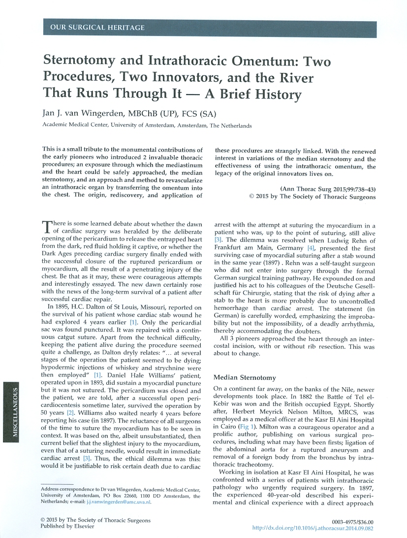
On on a continent far away, on the banks of the Nile, newer developments took place. In 1882 the Battle of Tel el-Kebir was won and the British occupied Egypt. Shortly after, Herbert Meyrick Nelson Milton, MRCS, was employed as a medical officer at the Kasr El Aini Hospital in Cairo.... He was confronted with a series of patients with intrathoracic pathology who urgently required surgery.
Median Sternotomy
In 1897, the experienced 40-year-old described his experimental and clinical experience with a direct approach to the mediastinum through what he termed an "anterior median thoracic incision", or what we would today call a sternotomy. Milton recognized the potential of his new exposure to the heart when he avowed: "So easy is this incision of execution and so considerable is the power of exploration thereby obtained that one is almost induced to hope that future experience may justify the application to it of the term 'normal thoracic incision'".

Contrary to popular belief that the median sternotomy was only reintroduced approximately half a century later, it remained a definite approach to the anterior mediastinum and its structures in the interim. For example, in 1929, in what they called their "final" report on all surgical cases with chronic valvular disease and the procedures, Elliott Cutler and Claude Beck from Boston, enthusiastically propounded "The medium sternotomy exposure was used in seven of ten patients operated on. An excellent exposure of the heart is obtained by this method and it also has the added advantage of not opening the pleura. They concluded that "wide exposure of the heart and approach to the valve through the ventricle is more desirable...for which a sternotomy is ideally suited, but also warn the uninitiated that "it is an extensive operation in itself...".
Omentum in Complex Cardiothoracic Problems
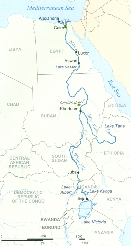
One year after the publication of Milton's seminal paper, in 1898, the brutal and bloody Battle of Omdurman was fought and won by the British. With the establishment of an Anglo-Egyptian administration a year later, British domination over Sudan became a reality. Omdurman lies close to where the exhausted White Nile is replenished by the more energetic Blue Nile.

It is here that one of the most dynamic figures in the history of thoracic surgery was to be found: Laurence O'Shaughnessy. Omdurman for O'Shaughnessy represented not only the place where 2 great rivers conjoined, but also the place where he became a proficient surgeon, developed a profound interest in thoracic surgery, and proceeded to learn German in his leisure time (to be able to visit Ferdinand Sauerbruch in Berlin during intervals of leave). Why Sauerbruch?
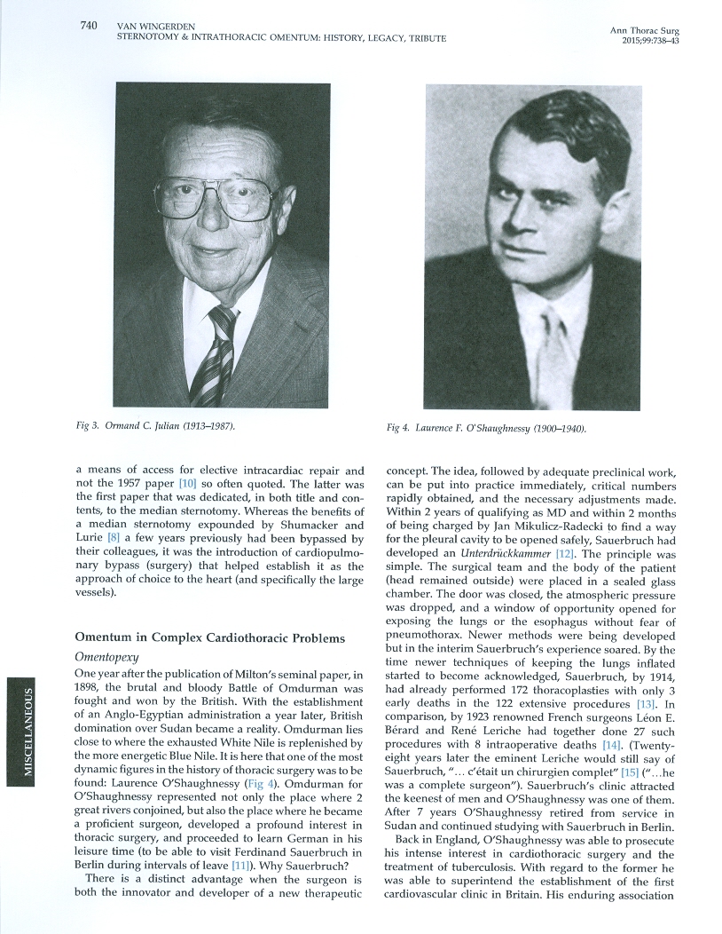
There is a distinct advantage when the surgeon is both the innovator and developer of a new therapeutic concept.... Within 2 years of qualifying as MD and within 2 months of being charged by Jan Mikulicz-Radecki to find a way for the pleural cavity to be opened safely, Sauerbruch had developed an Unterdruckkammer. The principle was simple. The surgical team and the body of the patient (head remained outside) were placed in a sealed glass chamber. The door was closed, the atmospheric pressure was dropped, and a window of opportunity opened for exposing the lungs or the esophagus without fear of pneumothorax.... Sauerbuch, by 1914, had already performed 172 thoracoplasties.... Sauerbruch's clinic attracted the keenest of men and O'Shaughnessy was one of them. After 7 years O'Shaughnessy retired from service in Sudan and continued studying with Sauerbruch in Berlin.
Back in England, O'Shaughnessy was able to prosecute his intense interest in cardiothoracic surgery and the treatment of tuberculosis. With regard to the former he was able to superintend the establishment of the first cardiovascular clinic in Britain. His enduring association with Sauerbruch culminated in the publication in 1937 of a revised and abridged English edition of Sauerbruch's Die Chirurgie der Brustorgane.
What set O'Shaughnessy apart was the difficulty of the problems he sought to solve, the vigor with which they were pursued, and the unique solutions he proposed. Up to then, all the focus regarding revascularization of tissue had been concentrated on vessel surgery (unblocking, bypassing, or modulating through the autonomic nervous system). Cardiac ischemia, however, remained a problem. O'Shaughnessy spawned the idea of bringing in tissue with excellent vascularization abilities and devised the route to get it to the ischemic heart. He chose omentum.
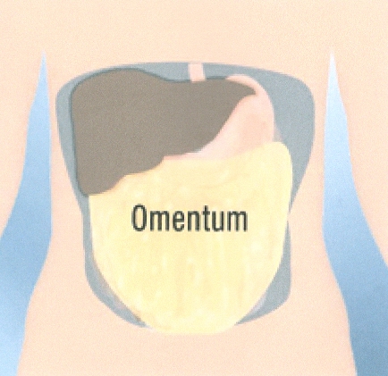
The idea of using omentum to increase myocardial perfusion germinated in Africa, for O'Shaughnessy attests: "Personal experience of it in the Sudan confirmed my belief in its value...". Apart from his own experience, he was well versed in the relevant literature; among others, he was familiar with the work of Lanz.
Otto Lanz of Amsterdam, former protege of E. Theodor Kocher, was the first to draw attention to the unique vascularization abilities of the omentum by wrapping it around denuded intestine in 1906. He concludes that "a vascularly threatened abdominal organ can be replenished by the omentum". He was later to clinically confirm the feat by simply attaching (instead of wrapping) the omentum to the completely devascularized colon, omentopexy.

The route to the heart chosen by O'Shaughnessy was through the diaphragm. By opening the diaphragm to allow the omentum to pass through, a sluice gate for increased vascular supply to the chest was unlocked. The omentum was sutured to the myocardium at the apex from where it could form attachments partly in the territories of the left and right coronary arteries. The technique became known as cardio-omentopexy. It was rapidly adopted outside England with the first case being recorded in the Netherlands in 1939 by Albert Kummer. By selecting the omentum, O'Shaughnessy had chosen tissue that was rich not only with vasculature but also with unique angiogenic ability. The neo-vascularization is beautifully illustrated in his classic paper. The angiogenic factors, such as vascular endothelial growth factor, with which the omentum is richly endowed, were only identified more than half a century later.
O'Shaughnessy died at the age of 39 years, on May 27, 1940 at Dunkirk. For a long time the circumstances at his death were shrouded in mystery. Were the story true, it would have been a wonderful testimony to enduring friendship and mutual respect, despite the change of circumstances that a war brings about. It has long been believed that O'Shaughnessy suffered a serious chest wound and that Sauerbruch had been granted a pass to cross British lines to come to the aid of his "disciple" (as O'Shaughnessy called himself). The former is unknown, the latter very unlikely. From the recently published war diary of Major George McNab, FRCP, a chest physician, who was with and attended to the mortally wounded O'Shaughnessy, we read that he died within half an hour after being struck by a bomb splinter. Time would have been too short to get word to the British superiors, let alone to Sauerbruch. Moreover, the exact nature of the injury suffered was never recorded and thus remained unknown to McNab's superiors and Sauerbruch.
Omentoplasty
A median sternotomy, not unlike other extensile exposures, can be complicated by severe mediastinitis. Although this was not frequent, mortality approached 50% in the early series.... The 3 surgeons reasoned that the infected cartilages were the main contributing factor to the post-sternotomy mediastinitis and a radical solution was found: the total excision of the sternum and adjacent costal cartilages and transposition of the (vascularized) omentum into the defect.... They were the first recorded cases of employing the omentum in the management of post-sternotomy mediastinitis. The concept was readily accepted and the introduction of flaps revolutionized the treatment of post-sternotomy mediastinitis, rapidly reducing the mortality.
Legacy
How exactly the omentum combats the infection is still poorly understood. Only recently has it been shown that upon stimulation the omentum acquires a large number of immunomodulatory cells. This is a characteristic quite unique to the omentum. It explains its steadily increasing popularity in the treatment of post-sternotomy mediastinitis.
In surgical treatment of post-sternotomy mediastinitis, the omentum was originally approached through a median laparotomy. More recently, it has been simplified by harvesting the omentum laparoscopically.... Moreover, previous abdominal surgery has been shown not to be a contraindication to laparoscopic harvesting of an omental flap.... The omentum is of particular benefit where significant sternal loss has occurred and bulk is required to fill the dead space. A clear preference has been expressed for the use of omentum (instead of muscle) in cases where the primary causative organism is particularly resistant, such as methicillin-resistant Staphylococcus aureus or Candida, or where the patient suffers from diabetes melitus.
A prerequisite for successful cardiac revascularization by means of coronary artery bypass grafting is the presence of a viable, albeit ischemic, myocardium. Once the myocardial damage has become so extensive that end-stage congestive heart failure is reached, only heart transplantation could offer hope. However, due to the limitations of donor organ availability and strict selection criteria, alternative methods of restoring myocardium were sought. Stem cell-based therapy entails finding the most effective method of specific, controlled cardiomyocyte regeneration by either replacing nonviable myocardium or improving the function, such as contractility, of the remaining and possibly recruited new cells throuth paracrine effects. The stem cells are either injected (also known as cellular cardiomyoplasty) or delivered to the injured cardiac region contained in cell sheets or scaffolds (cardiac tissue or organ engineering).
During translation from bench to bedside, it rapidly became clear that whichever method was chosen, only good blood supply would ensure the survival of the transplanted stem cells. Thus, after almost 60 years, the omentum was rediscovered as the ideal source for blood supply and, as additional benefits, as having angiogenic potential and regenerative properties. It is absolutely fascinating how much the more recent illustration of the new collaterals developed reminds one of similar illustrations in O'Shaughnessy's paper. The omentum is now regularly employed in furthering stem cell-based research for chronic ischemic heart failure and is starting to find clinical application as well.
Conclusions
"Eventually, all things merge into one, and a river runs through it", concludes the narrator in the beautiful book by the esteemed scholar and author Norman Maclean. It may be that modern cardiac surgery started near the confluence of 2 other big rivers (the Missouri and the Mississippi) at St Louis, was facilitated by surgical advancements on the Nile, and was developed further by ideas spawned near the confluence of 2 further great rivers (the Blue Nile and the White Nile). It is, however, another river that I was thinking of: in the words of David Brooks -- it is the river of knowledge we inherit and drink from with a flow of patterns that come from many sources that profoundly molds our thoughts. Innovators and discoverers contribute to the source. Our mentors added their knowledge and wisdom, and so the river continues to grow and flow. To them, we should be grateful.
~ end quoting Two Procedures/Two Innovators by van Wingerden ~
HOW ORWELL DOC O'SHAUGHNESSY DIED
ORWELL DOC O'SHAUGHNESSY GIANT IN FIELD
ORWELL BROTHER-IN-LAW DIED AT DUNKIRK
ORWELL'S HAMPSTEAD FLAT (met future wife Eileen O'Shaughnessy)
Ernst Ferdinand Sauerbruch (1875-1951) was a German surgeon. Sauerbruch was born in Barmen, Germany. He studied medicine at the Philipps University of Marburg, the University of Greifswald, the Friedrich Schiller University of Jena, and the University of Leipzig, from the last of which he graduated in 1902. He went to Breslau in 1903, where he developed the Sauerbruch chamber, a pressure chamber for operating on the open thorax, which he demonstrated in 1904. This invention was a breakthrough in thorax medicine and allowed heart and lung operations to take place at greatly reduced risk. As a battlefield surgeon during World War I, he developed several new types of limb prostheses, which for the first time enabled simple movements to be executed with the remaining muscle of the patient. Sauerbruch worked at the Ludwig Maximilians University of Munich from 1918 to 1927 on surgical techniques and diets for treating tuberculosis. From 1928 to 1949, he was the head of the surgical department at the Charite in Berlin, attaining international fame for his innovative operations. Because of his experience and extraordinary skills he quickly attained an international reputation and operated on many prominent patients. At the same time he was well known for his uncompromising and passionate dedication to all patients independent of their social, political or ethnical backgrounds. Before World War II, the Nazi Government awarded him the German National Prize for Art and Science. Sauerbruch's position towards the Nazi government is ambiguous and the subject of debate. In his position he was clearly in contact with the political elite but he was never a member of and did not support the political objectives of the NSDAP. He was, however, a fervent nationalist who wanted to undo the "humiliation of Versailles" and was keen to show off his country as an advanced and sophisticated society....
Cardiac arrest happens when your heart stops pumping blood around your body.
Cardio-omentopoxy is surgical attachment of the omentum to the heart to improve its blood supply.
Congestive heart failure (CHF) is a condition in which the heart's function as a pump is inadequate to meet the body's needs.
Ischemia is a restriction in blood supply to tissues, causing a shortage of oxygen and glucose needed for cellular metabolism (to keep tissue alive). Ischemia is generally caused by problems with blood vessels, with resultant damage to or dysfunction of tissue.
Intercostal means "between the ribs" of an animal or person
Intrathoracic means within the thorax or chest
Laparoscopic surgery, also called minimally invasive surgery (MIS), bandaid surgery, or keyhole surgery, is a modern surgical technique in which operations are performed far from their location through small incisions (usually 0.5-1.5 cm) elsewhere in the body. There are a number of advantages to the patient with laparoscopic surgery versus the more common, open procedure. Pain and hemorrhaging are reduced due to smaller incisions and recovery times are shorter. The key element in laparoscopic surgery is the use of a laparoscope, a long fiber optic cable system which allows viewing of the affected area by snaking the cable from a more distant, but more easily accessible location.
Median sternotomy is a type of surgical procedure in which a vertical inline incision is made along the sternum, after which the sternum itself is divided, or "cracked". This procedure provides access to the heart and lungs for surgical procedures such as heart transplant, corrective surgery for congenital heart defects, or coronary artery bypass surgery.
Mediastinitis is inflammation of the mediastinum. Acute mediastinitis usually results from esophageal perforation or median sternotomy. Symptoms include severe chest pain, dyspnea, and fever. The diagnosis is confirmed by chest x-ray or CT.
Mediastinum is the central compartment of the thoracic cavity surrounded by loose connective tissue, as an undelineated region that contains a group of structures within the thorax. The mediastinum contains the heart and its vessels, the esophagus, trachea, phrenic and cardiac nerves, the thoracic duct, thymus and lymph nodes of the central chest.
Myocardial infarction (MI), commonly known as a heart attack, occurs when blood stops flowing properly to a part of the heart, and the heart muscle is injured because it is not receiving enough oxygen. Usually, this is because one of the coronary arteries that supplies blood to the heart develops a blockage due to an unstable buildup of white blood cells, cholesterol and fat. The event is called "acute" if it is sudden and serious. Myocardial infarction differs from cardiac arrest, although cardiac arrest can be a consequence of MI.
Myocardial ischemia occurs when blood flow to your heart muscle is decreased by a partial or complete blockage of your heart's arteries (coronary arteries). The decrease in blood flow reduces your heart's oxygen supply. Myocardial ischemia, also called cardiac ischemia, can damage your heart muscle, reducing its ability to pump efficiently. A sudden, severe blockage of a coronary artery may lead to a heart attack. Myocardial ischemia may also cause serious abnormal heart rhythms. Treatment for myocardial ischemia is directed at improving blood flow to the heart muscle and may include medications, a procedure to open blocked arteries or coronary artery bypass surgery. Making heart-healthy lifestyle choices is important in treating and preventing myocardial ischemia.
Myocardium is the muscular tissue of the heart. Other tissues are the endocardium (inner lining, effectively a specialised endothelium) and the pericardium (a connective tissue layer around the heart). The myocardium is the middle layer of the walls of the heart, made of cardiac muscle, that contracts to push out blood. The heart muscle can become sick and weak. Heart muscle can even die if blood flow stops. Most problems with the heart muscle come from defects in the blood supply. The regular contractions of the heart need an unending supply of oxygen from the arteries supplying the heart muscle.
Omen (also called portent or presage) is a phenomenon that is believed to foretell the future, often signifying the advent of change. The origin of the word is unknown, although it may be connected with the Latin word audire, meaning "to hear".
Omentoplasty is a surgical procedure in which part of the greater omentum is used to cover or fill a defect, augment arterial or portal venous circulation, absorb effusions, or increase lymphatic drainage. The omentum has been described as the "policeman of the abdomen", in that it wraps around abdominal structures such as the gallbladder and appendix and can revascularize them when they are deprived of their blood supply.
Omentum is a part of the body that is found in your lower abdominal area. It is made up of two layers of fatty tissues and both supports and covers the organs and intestines found in this area of the body. There are two parts of the omentum, the greater omentum and the lesser omentum, which are responsible for storing fat deposits and connecting the intestines and stomach to the liver respectively.
Pericardial Sac (from the Greek "around" and "heart") is a double-walled sac containing the heart and the roots of the great vessels. The pericardial sac has two layers, a serous layer and a fibrous layer. It encloses the pericardial cavity which contains pericardial fluid. The pericardium fixes the heart to the mediastinum, gives protection against infection, and provides the lubrication for the heart.
Pleura is one of the two membranes around the lungs. These two membranes are called the visceral and parietal pleurae. The visceral pleura envelops the lung, and the parietal pleura lines the inner chest wall.
Pleural cavity is the space that lies between the pleura, the two thin membranes that line and surround the lungs. The pleural cavity contains a small amount of a thin fluid known as the pleural fluid, which provides lubrication as the lungs expand and contract during respiration.
Pneumothorax is air that is trapped between a lung and the chest wall. The air gets there either from the lungs or from outside the body.
Sternum or breastbone is a long flat bony plate shaped like a capital "T" located anteriorly to the heart in the center of the thorax (chest). It connects to the rib bones via cartilage, forming the anterior section of the rib cage with them, and thus helps to protect the lungs, heart and major blood vessels from physical trauma. Although it is fused, the sternum can be sub-divided into three regions: the manubrium, the body, and the xiphoid process....
Thoracic surgery is the branch of surgery that treats the structures of the chest with the exception of the heart. Also known as: chest surgery, lung surgery. A thoracic surgeon is trained in the surgical treatment of the esophagus, lungs, chest muscle and the diaphragm muscle. A cardiothoracic surgeon is a thoracic surgeon also trained in surgery performed on the heart.
Thoracoplasty is the operation removing selected portions of the ribs to collapse part of the underlying lung or an abnormal pleural space, usually in the treatment of tuberculosis.
Thorax or chest is a part of the anatomy of humans and various other animals located between the neck and the abdomen. The thorax includes the thoracic cavity and the thoracic wall. It contains organs including the heart, lungs and thymus gland, as well as muscles and various other internal structures. Many diseases may affect the chest, and one of the most common symptoms is chest pain....
Tuberculosis (TB) is a potentially fatal contagious disease that can affect almost any part of the body but is mainly an infection of the lungs. It is caused by a bacterial microorganism, the tubercle bacillus or Mycobacterium tuberculosis. Although TB can be treated, cured, and can be prevented if persons at risk take certain drugs, scientists have never come close to wiping it out. Few diseases have caused so much distressing illness for centuries and claimed so many lives.
Vascularization (Re-vascularization) is the restoration of the blood circulation of an organ or area, achieved by unblocking obstructed or disrupted blood vessels or by surgically implanting replacements.
ORWELL DOC O'SHAUGHNESSY HEART OMEN
(contributor to source of river of knowledge)
Annals Thoracic Surgery, Feb 15, 2015
To Orwell Today,
Thank you for sharing and well done!
-Jan J van Wingerden
February 2015
Greetings Jan van Wingerden,
I just posted a new article about the death of Dr Laurence O'Shaughnessy you will no doubt be interested in reading:
ORWELL'S DOC O'SHAUGHNESSY'S GRAVE

(remembered with honour at Dunkirk Memorial)
Email, Aug 2, 2019
I also want to tell you that I've watched the movie A RIVER RUNS THRUOUGH IT which you use as a metaphor in your paper about the surgical heritage of O'Shaughnessy's contributions to your field of study.


I've also read a book of the other writings of Norman Maclean -- including his unfinished manuscript on the life of General George Armstrong Custer and the Battle of Little Bighorn -- the area of the Missouri and Yellowstone rivers around where Maclean fished. I, being an American Civil War and Indian Wars buff, am also researching the fascinating life and times of Custer.
All the best,
Jackie Jura, August 2019
Jackie Jura
~ an independent researcher monitoring local, national and international events ~
email: orwelltoday@gmail.com
HOME PAGE
website: www.orwelltoday.com